

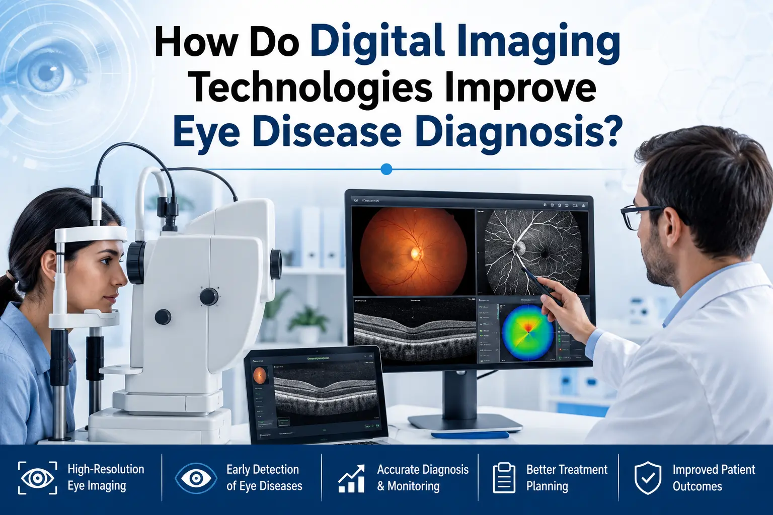
Digital imaging technologies help doctors see the eye in much more detail. Instead of depending only on symptoms, the doctor can study images of the retina, cornea, optic nerve and blood vessels.
In an eye hospital, these reports help detect disease early, confirm the cause of vision changes and decide the right treatment with better clarity. This is useful for patients who may delay checkups until discomfort becomes serious.
1. Early Signs Become Easier to Detect
Many eye diseases develop gradually. A person may not feel pain or notice vision loss in the early stage. Digital imaging can show small changes such as retinal swelling, tiny blood leaks, optic nerve thinning or changes in the cornea. These findings help doctors identify a condition before it starts affecting vision more seriously.
2. Deeper Eye Structures Become Clearer
A regular eye checkup is important, but some parts of the eye need detailed scanning. OCT scans show the layers of the retina and optic nerve. Fundus photography captures the back of the eye. Angiography helps doctors check blood flow in the retina. These images help an eye hospital understand where the problem is and how much of the eye is affected.
3. Diagnosis Becomes More Accurate
Blurred vision, redness, headache or eye strain can happen due to different reasons. Digital imaging helps doctors separate one condition from another. For instance, blurred vision may be due to spectacle power, cataract, retinal swelling or nerve-related changes. When scan findings are studied with eye pressure, vision tests and medical history, the diagnosis becomes more reliable.
4. Treatment Planning Becomes Easier
Clear images help doctors decide whether the patient needs medicines, laser treatment, injections, surgery or only regular monitoring. The report also helps explain how urgent the condition is. This makes the treatment process easier for patients and families to understand. In an eye care hospital, imaging reports often support safer and more practical treatment decisions.
5. Follow-up Visits Show Real Progress
Some eye conditions need regular follow-up, especially diabetes related eye disease, glaucoma and retinal problems. Digital imaging allows doctors to compare old and new reports. If swelling has reduced or nerve changes are stable, it can be seen clearly. If the condition is progressing, treatment can be changed at the right time.
6. Choosing Care Becomes More Sensible
When searching for an eye hospital near me, patients should check more than location. The best eye hospital should have trained specialists, diagnostic support and clear reporting. Good testing facilities help doctors study the condition properly before advising treatment, especially when symptoms are unclear or long-lasting.
Final Thoughts
Digital imaging helps doctors spot eye issues earlier and understand them better. It gives a much clearer view of what is happening inside the eye, so diagnosis, treatment and even follow-up become more precise. A timely visit to an eye hospital can help patients to receive the proper guidance before the condition becomes more difficult to manage.
FAQs
1. How does digital imaging help in eye diagnosis?
It gives the doctor a clearer view inside the eye, so small problems can be found before they become serious.
2. Who may need digital eye imaging?
People with diabetes, glaucoma risk, retina concerns, sudden vision changes or long-term eye discomfort may need imaging after the doctor’s advice.
3. Can imaging reports help during follow-up visits?
Yes, old and new images help the doctor understand whether the condition is improving, stable or getting worse.
4. Is digital imaging done for every patient?
Not always. Imaging is usually advised only when the doctor needs a closer look at the retina, cornea or optic nerve before deciding the next step.